 |
||
|
||
| ||
|
||
|
||
| ||
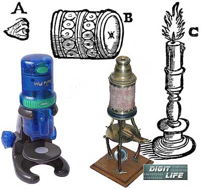 One of the earliest forms of a simple microscope was initially described by Athanasius Kircher in 1646. As a toy it was widespread up to the end of XVIII century. On the one end of a cardboard tube they mounted a biconvex lens, and on the other end there was flat glass with an object attached to it. It's considered that Nuremberg craftsmen made their cheap cardboard-wooden, simple and complicated microscopes covered with color or black paper around 1750. Note that a toy microscope was a still working instrument. And now, two and a half centuries later, Intel keeps the traditions of toy microscope production. And like Nurmberg craftsmen who decorated them with red or green chest-board pattern, Intel paid special attention to the loud decoration of their toys. The semiconductor body of a new Intel Play QX3 microscope is far from functionality and is implemented in a modern style of iMAC. Despite its a 3-century history the microscope has become nearly the most popular and famous optical device. Its development continued in the XX century as well. In 1953 Frits Zernike was awarded the Nobel Prize for a phase-contrast microscope. You understand that the toy is not going to compete against modern scientific microscopes, but it has a lot of competitors in its class. That's why there is something to compare with. In this article we will compare possibilities of the microscope in question and results obtained when using a child microscope "Naturalist", a Carl Zeiss microscope of the beginning of the century and a usual scanner which can also be used to receive an enlarged photo of rather small objects. 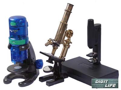 This toy is aimed at receiving photos of minute objects and not intended for direct viewing of them. That's why for correct comparison we carried out shooting through the optical microscope with different digital and video cameras. Here are an optical scheme of the microscope and main shooting methods through it. Optical schematics of a microscope:  a) visual examination 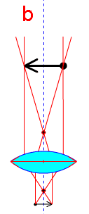 b) photomicrography with a lens of the microscope 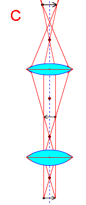 c) photomicrography with an ocular 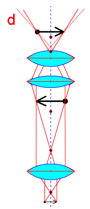 d) photomicrography with the lens of the photo camera 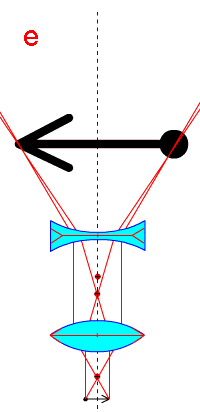 e) photomicrography with a gammal The pictures above demonstrate optical schematics of the microscope: a) visual examination (the oldest scheme of a biconvex lens and plano-convex ocular , is considered to be suggested by Drebbel in 1617-1619), b) photomicrography with a lens of the microscope, c) photomicrography with an ocular, d) photomicrography with the lens of the photo camera, e) photomicrography with a gammal. In the Intel Play QX3 microscope the b) variant is used. Since the majority of cheap digital photo and video cameras has the lens stationary, the variant d) is the most convenient when shooting through a usual microscope. When using a camera with a stationary lens for photomicrography the ocular becomes the most important element for obtaining high quality image. If a primary image created with the microscope lens is a bit higher than a front focus of the ocular, then there will be divergent beams coming out of it. Running into a positive optical system (the camera's lens) they will create an image a bit farther than a main focal plane of this system. The microscope plays a role of an afocal attachment to the photo camera. That's why let's consider the main types of oculars.  a) Huygens's ocular , b) compensating ocular, c) Ramsden's ocular, d) orthoscopic ocular , e) symmetrical ocular , f) gammal. The most interesting for us is the symmetrical ocular e). It consists of two identical symmetrically positioned lenses of the same diameter. Each lens is achromatic and sewn of two elements. Its optical construction is much similar to the construction of many standard photographic lenses which can be used instead of the ocular . Now come the objects under examination. The micropreparations were made as cardboard plates with 4 round windows where the objects were located between two transparent plates. They were: a louse, a bit of a pheasant feather, a leg and a wing of a bee, wool, fruit-fly's body, shrimp's berries, a fragment of a sponge (of an invertebrate), a thread of Spirogyra, spores of a fern, fibers of paper and a bit of dog's hair. "Leg of a bee"  NEUHAUS 9600 scanner 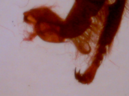 Intel Play QX3 (the whole still) 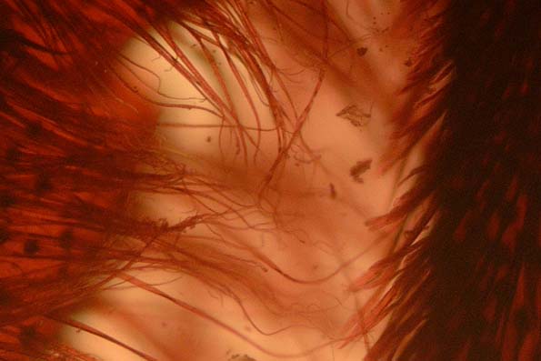 FujiFilm FinePix 4700 + Carl Zeiss microscope (a fragment) What we have received you can see on these photos, and the shots made with the Naturalist and Carl Zeiss microscopes with Huygens oculars and with the help of the Fuji cameras are printed in the article FujiFilm FinePix 4700. The shots show that having added such feature as 200x the designers of the microscope forgot to correspond it with the number of pixels.  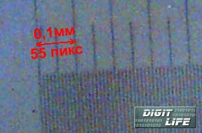 The resolving capacity of a human eye is about 0.1 mm. That's why, in order to make a comparison of magnification of the optical microscope and our device you need to print a shot at resolution of 10 pixels per mm or 254 dpi on a sublimation printer which provides a distance between the neighbouring pixels to be less than 0.1 mm. The object 's size on the imprint related to its original size will give us the desired magnification. So, the image of the object 0.1 mm in size consists of 55 pixels and will take 5.5 mm on the print; it means that the magnification is 55. The microscope is supplied with three lenses. The magnification of the weakest one seems to be less than 1, since the size of the objects is several times more than the assumed matrix's size. I think that the scheme with 3 lenses is not justified here. A gradual magnification change at the expense of the distance change between an object and the matrix would allow the same range of magnification. An obvious advantage of weak lenses is a huge distance between an object and a lens what allows effective upper highlight. The Intel microscope is supplied with two lighters allowing to direct the light both from above and below. Here is used the simplest lighter consisting of a bulb and opal glass. 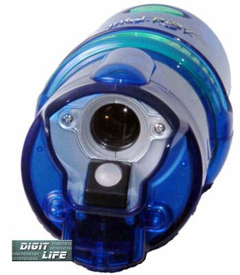  Our hero is next to the Levenhook's microscope. In sharpening and magnification it falls behind. A short-focus lens in a case, connected with a tripod mounting for fixing an object and focusing became a scientific tool in the hands of Levenhook. Its advantages over complex microscopes of that time were so obvious that it for a long time served a basic instrument for all good microscopists. That's why the main attention in microscope construction was paid mainly to the mechanical part improvement. The mechanics of our microscope is hardly comes later than that of the XVIII century. What's good, then, in our microscope? It allows viewing the image by several observers simultaneously in real time. And in very comfortable and convenient position sitting in front a display of the computer, and not projecting an image on the ceiling in the dark room. Besides, there is a possibility to fix an object at any moment, add writings and ... retouch and paint it with the help of the supplied software. This program immediately sets the 800 X 600 mode where in the 512 X 384 window you can see an object, and the remaining part is filled up with buttons for microscope control and image processing.  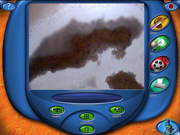
The device must be called for, and here you can read impressions of an unprejudiced observer. First time I heard about a possibility to view the image created by a microscope on the display somewhere in 80s. It was at the department of microbiology of the Sverdlovsk University. I stared at the screen and envied. The impression was very strong. Ten years later we combined a microscope with an inserted lighter, a black-&-white video camera and a monitor, it allowed showing the preparation to all students at the same time being sure that all of them saw the same and did not focus different planes of the preparation. Especially convenient was a construction for observing living objects (bacteria, protozoa, alga etc.), observing movements of microorganisms, process of getting and swallowing the food etc. We made an expedition while we were showing to different people the life in a drop if water. Everybody was greatly excited. The second impression: an attempt to examine customary (and the largest) objects for a microbiologist - tiny alga, cyanobacteria etc. resulted in disappointment. Apart from indistinct images we could see nothing. The same result was achieved with Rotifera - microscopical parasites of a crab. It was clearly distinct in a usual microscope but became an unclear cloud when observing it on the screen. The third impression: we were looking at everything we could reach. First of all they were minute insects, legs, wings, berries which are hundreds times larger the chosen microbes. Here the situation was much better - of course it was not ideal, but we still could focus and examine the objects. Here I'd like to say some words about the suggested preparations. The set was quite standard. Obviously unsuccessful was a preparation of the Spirogyra, it is alga with rather gross threads and noticeable cellular membranes and bright green spiral chromatophore. On the preparation the chromatophore was not seen, thread were colorless and formless. In other respects, the preparations were made acceptable (despite the fact that the Latin names were lacking). Besides, the obtained images on the display looked a bit different than when looking into the light microscope. But it was rather curious. The most interesting object to examine turned to be rather big objects at hand: beautiful jewelry, stones, cactus needles, a cut of a leaf. It seems that these objects, which could be rather clearly seen with the naked eye, turned to be the best for this microscope: on the display they change their images, get rich of new details and stimulate intellectual curiosity. In this relation there is a good possibility of top highlight. That's why the impression is optimistic. 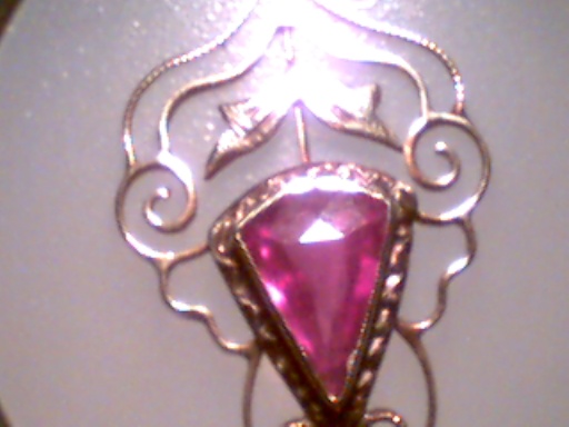 The forth impression. The microscope is detachable and you can take off the microscope from the base and take it to any object, let it be seat upholstery, window glass etc. That's why its possibilities are rather wide.  The fifth impression. An absolute advantage of
the toy is a possibility of a simultaneous joint examination, a
joint game and joint art since you can not only see the object but
also paint it, change it, make a composition, add a writing. Well,
as an optical instrument it has a lot of downsides, but on the other
hand it is a beautiful, fascinating, educating toy.
Write a comment below. No registration needed!
|
Platform · Video · Multimedia · Mobile · Other || About us & Privacy policy · Twitter · Facebook Copyright © Byrds Research & Publishing, Ltd., 1997–2011. All rights reserved. |